Upper Leg Tendon Anatomy : Simple Diagram Of Leg Muscles Largest Wiring Diagram Database - Upper legs anatomy — stock image.
Upper Leg Tendon Anatomy : Simple Diagram Of Leg Muscles Largest Wiring Diagram Database - Upper legs anatomy — stock image.. Palmar region , arteries (illustrations: 630 anatomical structures of the upper limb (pectoral girdle, shoulder, arm, elbow, forearm, wrist, hand and fingers) were labeled. To describe the mechanical properties of tendons. The tendons that control movement in your hands, wrists and fingers run through your forearm. There are four muscles in the anterior compartment of the leg.
Legs come in all shapes and sizes, ranging from portly and stout, to the streamlined, almost emaciated legs of runway models, to the muscular legs of athletes. To download this image, create an account. Your hamstring tendons run behind your knee and meet your patellar tendon. The upper leg begins at the hip and continues down to the knee. The thigh bone, or femur, is the large upper leg bone that connects the lower leg bones (knee joint) to the pelvic bone (hip joint).
Muscle Anatomy from droualb.faculty.mjc.edu The thigh bone, or femur, is the large upper leg bone that connects the lower leg bones (knee joint) to the pelvic bone (hip joint). The human leg, in the general word sense, is the entire lower limb of the human body, including the foot, thigh and even the hip or gluteal region. When tendons become inflamed, irritated or suffer microscopic tears, the condition is called tendonitis. Artists usually begin their study of the legs by. This mri wrist coronal cross sectional anatomy tool is absolutely free to use. Try this movement out by standing on one foot with the other leg. To describe the mechanical properties of tendons. Fibula— a long, thin bone in the lower leg on the lateral side which runs along side the tibia from the knee to the ankle.
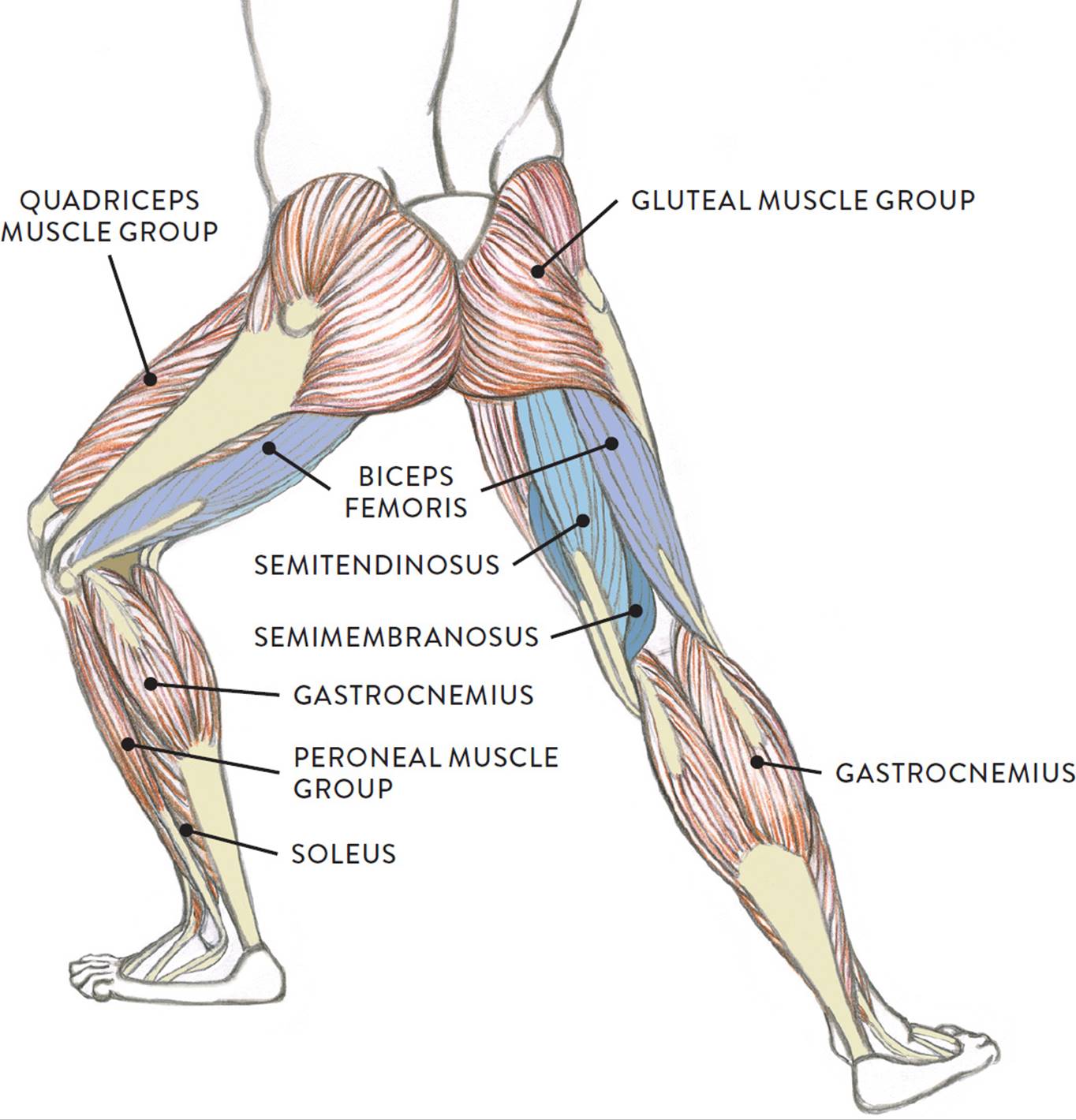
The leg anatomy includes the quads, hams, glutes, hip flexors, adductors & abductors.
Palmar region , arteries (illustrations: Learn the origin/insertion, functions & exercises for the leg rotating your upper leg and pelvis to the inside or outside of your body's center line. When tendons become inflamed, irritated or suffer microscopic tears, the condition is called tendonitis. Learn vocabulary, terms and more with flashcards, games and other study tools. Related online courses on physioplus. Leg anatomy anatomy poses anatomy study anatomy art anatomy drawing human anatomy anatomy images body reference anatomy anatomical drawings sketchbook ,artist study resources for art students with thanks to artist simone bianchi, how to draw the human figure. Lateral supracondylar line of femur, oblique popliteal ligament of knee insertion: The leg anatomy includes the quads, hams, glutes, hip flexors, adductors & abductors. To download this image, create an account. The human leg, in the general word sense, is the entire lower limb of the human body, including the foot, thigh and even the hip or gluteal region. Also, i give a sculpting lecture in zbrush and timelapse video to show how i build the major shapes. Collectively, they act to dorsiflex and invert the foot at the ankle joint. If you tear your biceps tendon at the shoulder, you may lose some strength in your arm and have pain when you forcefully turn your arm from palm down to palm up.
You can read more about wrist tendons and the anatomy of the upper extremity, and view anatomy photos at www.handcare.org. Upper legs anatomy — stock image. Tendinous sheath of right flexor pollicis longus radial bursa. Upper leg anatomy and function. Want to learn more about it?
Muscles Of The Leg And Foot Classic Human Anatomy In Motion The Artist S Guide To The Dynamics Of Figure Drawing from doctorlib.info Palmar region , arteries (illustrations: Leg anatomy anatomy poses anatomy study anatomy art anatomy drawing human anatomy anatomy images body reference anatomy anatomical drawings sketchbook ,artist study resources for art students with thanks to artist simone bianchi, how to draw the human figure. Tendons can be small, like the delicate, tiny bands in the hands, or large, like the heavy, ropelike cords that anchor the calf or thigh muscles. Upper leg, knee, lower leg, ankle, and foot. Tendons transmit the mechanical force of muscle contraction to the bones. Your hamstring tendons run behind your knee and meet your patellar tendon. This mri wrist coronal cross sectional anatomy tool is absolutely free to use. Movement at the hip joint occurs when you tendons that help you bend or straighten the knee include:
The nerve signals in these reflexes come from stretch receptors located in the joints, ligaments reflexes help to maintain proper muscle tone, balance, and responsiveness of the legs and feet to stimuli such as stepping on a sharp object.
Tendon, tissue that attaches a muscle to other body parts, usually bones. The upper leg begins at the hip and continues down to the knee. The thigh bone, or femur, is the large upper leg bone that connects the lower leg bones (knee joint) to the pelvic bone (hip joint). Synovial tendon sheaths of right fingers. To download this image, create an account. Upper limb trauma programme injuries. The human leg, in the general word sense, is the entire lower limb of the human body, including the foot, thigh and even the hip or gluteal region. Learn the origin/insertion, functions & exercises for the leg rotating your upper leg and pelvis to the inside or outside of your body's center line. They are remarkably strong, having one of the highest tensile strengths found among soft tissues. Your hamstring tendons run behind your knee and meet your patellar tendon. Tendinous sheath of right flexor pollicis longus radial bursa. Movement at the hip joint occurs when you tendons that help you bend or straighten the knee include: Learn vocabulary, terms and more with flashcards, games and other study tools.
Your biceps tendons attach the biceps muscle to bones in your shoulder and in your elbow. However, many reflex pathways are also active in the legs and foot. The upper leg begins at the hip and continues down to the knee. Quadriceps tendon to base of patella and onto tibial tuberosity via the patellar ligament action: The nerve signals in these reflexes come from stretch receptors located in the joints, ligaments reflexes help to maintain proper muscle tone, balance, and responsiveness of the legs and feet to stimuli such as stepping on a sharp object.
Muscular Function And Anatomy Of The Upper Leg Video Lesson Transcript Study Com from study.com To describe the mechanical properties of tendons. Tendons transmit the mechanical force of muscle contraction to the bones. A tendon is the fibrous tissue that attaches muscle to bone in the human body. Tendinous sheath of right flexor pollicis longus radial bursa. Learn the origin/insertion, functions & exercises for the leg rotating your upper leg and pelvis to the inside or outside of your body's center line. Learn vocabulary, terms and more with flashcards, games and other study tools. The leg is composed of five distinct sections: Posterior surface of calcaneus (via calcaneal tendon).
Legs come in all shapes and sizes, ranging from portly and stout, to the streamlined, almost emaciated legs of runway models, to the muscular legs of athletes.
Upper leg, knee, lower leg, ankle, and foot. Superficial veins of upper limb , anatomy : When tendons become inflamed, irritated or suffer microscopic tears, the condition is called tendonitis. There are four muscles in the anterior compartment of the leg. Artists usually begin their study of the legs by. Synovial tendon sheaths of right fingers. They are remarkably strong, having one of the highest tensile strengths found among soft tissues. The upper leg begins at the hip and continues down to the knee. Posterior surface of calcaneus (via calcaneal tendon). Collectively, they act to dorsiflex and invert the foot at the ankle joint. Quadriceps tendon to base of patella and onto tibial tuberosity via the patellar ligament action: The peroneus longus originates at the head of your fibula and the upper half of the shaft of your fibula on the outer part of your lower leg. Movement at the hip joint occurs when you tendons that help you bend or straighten the knee include: