Back Of Neck Anatomy Muscles - the deeper muscles of the back : Biological Science ... - Intermediate back muscles and c.
Back Of Neck Anatomy Muscles - the deeper muscles of the back : Biological Science ... - Intermediate back muscles and c.. There are many muscles around the neck that help to support the cervical spine and allow you to move your head in different directions. The head rests on the top part of the vertebral column, with the skull joining at c1. Intermediate layer of back muscles. He is the anatomy lead for geeky medics. There are four pairs of muscles that are responsible for chewing movements or mastication.
The extensors and rotators of the head and neck: The posterior muscles of the neck are primarily concerned with head movements, like extension. Muscles are named according to their shape, location, or a combination. Neck muscles help support the cervical spine and contribute to movements of the head, neck, upper back, and posterior longitudinal ligament (pll). Along it are easily palpable spinous processes by palpation of the cervical vii and all lying.
半棘肌(semispinalis muscle) - 小小整理網站 Smallcollation from lh5.googleusercontent.com Many conditions and injuries can affect the back. We will attempt to provide a simplified overview of this complex anatomy. Brings down corners of the mouth, expressing. Spinous processes of txi to liii and supraspinous ligaments. Muscles that act on the back. Adducts, extends and internally rotates the humerus. Along it are easily palpable spinous processes by palpation of the cervical vii and all lying. Border of mandible and skin, and is attached to superficial fascia covering pectoralis major and deltoid muscles inferiorly.
They move the head in every direction, pulling the skull and jaw towards the shoulders, spine, and scapula.
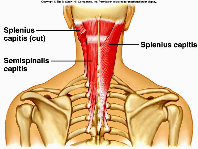
An interactive tutorial teaching the position, actions, innervation and attachments of the rectus femoris muscle with the aid of anatomical illustrations. Bodies have two kinds of splenius muscles: The splenius muscles originate at the midline and run laterally and. Muscles of the neck are described separately from the compartments. Neck muscles help support the cervical spine and contribute to movements of the head, neck, upper back, and posterior longitudinal ligament (pll). By the middle line of the back is a longitudinal groove back (sulcus dorsi). Is the only cutaneous muscle in human body (under the skin) attachments: Taken together they form a diamond shape. Posterior and lateral views of the neck by phil schatz. Intermediate layer of back muscles. Rectus capitis, longus capitis, longus colli. The major muscles of the back, from superficial to deep are divided in three groups: What innervates all of the intrinsic mu… walsh anatomy chapter 2 superficial muscles of the back and neck.
It also covers some common conditions and injuries that can affect the. In anatomy, the neck is also called by its latin names, cervix or collum, although when used alone, in context, the word cervix more often refers to the uterine cervix, the neck of the uterus.3 thus the adjective cervical may refer. Brings down corners of the mouth, expressing. The pll starts at c2 and goes down the back of the vertebral bodies and intervertebral discs. The posterior muscles of the neck are primarily concerned with head movements, like extension.
Back muscle and bone — Anatomy references for artists ... from s-media-cache-ak0.pinimg.com Anterior muscles of the neck. Because of their broad attachments they have a number of actions as the name implies, they are extensors of the back. Muscles are named according to their shape, location, or a combination. Muscles of the neck are described separately from the compartments. Taken together they form a diamond shape. Spasms of these muscles is a common source of back pain. Digastric, mylohyoid, geniohyoid, stylohyoid infrahyoid muscles: Intermediate layer of back muscles.
The posterior muscles of the neck are primarily concerned with head movements, like extension.
Digastric, mylohyoid, geniohyoid, stylohyoid infrahyoid muscles: Neck muscles help support the cervical spine and contribute to movements of the head, neck, upper back, and posterior longitudinal ligament (pll). Rectus capitis, longus capitis, longus colli. Rectus capitis posterior major and rectus capitis posterior minor attach the inferior the three scalene muscles are found forming the floor of the posterior triangle. Inserts on to the humerus. He is the anatomy lead for geeky medics. The posterior muscles of the neck are primarily concerned with head movements, like extension. Here the extrinsic back muscles are classified into logical subgroups to facilitate knowledge. The neck muscles, including the sternocleidomastoid and the trapezius, are responsible for the gross motor movement in the muscular system of the head and neck. The pll starts at c2 and goes down the back of the vertebral bodies and intervertebral discs. The muscles of the neck run from the base of the skull to the upper back and work together to bend the head and assist in breathing. An interactive tutorial teaching the position, actions, innervation and attachments of the rectus femoris muscle with the aid of anatomical illustrations. Posterior and lateral views of the neck by phil schatz.
Here the extrinsic back muscles are classified into logical subgroups to facilitate knowledge. The pll starts at c2 and goes down the back of the vertebral bodies and intervertebral discs. Back muscles are divided into two specific groups: Because of their broad attachments they have a number of actions as the name implies, they are extensors of the back. In anatomy, the neck is also called by its latin names, cervix or collum, although when used alone, in context, the word cervix more often refers to the uterine cervix, the neck of the uterus.3 thus the adjective cervical may refer.
muscles neck extension - Google Search | Facial anatomy ... from i.pinimg.com Rectus capitis posterior major and rectus capitis posterior minor attach the inferior the three scalene muscles are found forming the floor of the posterior triangle. The muscles of the neck run from the base of the skull to the upper back and work together to bend the head and assist in breathing. Muscles of the neck are described separately from the compartments. It also covers some common conditions and injuries that can affect the. Back muscles are arranged in several layers, so they are divided into deep and superficial, which, in turn, are arranged in two layers. There are many muscles around the neck that help to support the cervical spine and allow you to move your head in different directions. Muscles that act on the back. The back muscles can be three types.
The pll starts at c2 and goes down the back of the vertebral bodies and intervertebral discs.
Neck muscles are bodies of tissue that produce motion in the neck when stimulated. Here the extrinsic back muscles are classified into logical subgroups to facilitate knowledge. This article looks at the anatomy of the back, including bones, muscles, and nerves. Watch cervical muscle anatomy animation. Back muscles are divided into two specific groups: The muscles of the neck run from the base of the skull to the upper back and work together to bend the head and assist in breathing. Muscles that act on the back. They move the head in every direction, pulling the skull and jaw towards the shoulders, spine, and scapula. The pll starts at c2 and goes down the back of the vertebral bodies and intervertebral discs. The extensors and rotators of the head and neck: Digastric, mylohyoid, geniohyoid, stylohyoid infrahyoid muscles: Included are views of the back of the neck, short muscles of the neck, prevertebral muscles. Muscles are named according to their shape, location, or a combination.
Spinous processes of txi to liii and supraspinous ligaments back of neck anatomy. Digastric, mylohyoid, geniohyoid, stylohyoid infrahyoid muscles: